Home
/ Abdominal Anatomy Diagram - Organ Human Body Anatomy Torso Homo Sapiens Png Clipart Abdomen Anatomy Chest Finger Function Free Png : Gsi asked questions about the abdominal membranes to christopher windham, m.d.
Abdominal Anatomy Diagram - Organ Human Body Anatomy Torso Homo Sapiens Png Clipart Abdomen Anatomy Chest Finger Function Free Png : Gsi asked questions about the abdominal membranes to christopher windham, m.d.
Abdominal Anatomy Diagram - Organ Human Body Anatomy Torso Homo Sapiens Png Clipart Abdomen Anatomy Chest Finger Function Free Png : Gsi asked questions about the abdominal membranes to christopher windham, m.d.. A good amount of area is covered by the abdominal wall. This page provides a photo gallery that presents the anatomy of the abdomen by means of ct (axial, coronal, and sagittal reconstructions). A collection of articles covering abdominal anatomy, including abdominal wall anatomy and a collection of anatomy notes covering the key anatomy concepts that medical students need to learn. This article covers the anatomy of the rectus abdominis and pyramidalis muscles, their functions, and clinical anterior abdominal muscles: There are multiple anatomical areas within the abdomen, each of which contain specific contents and are bound by certain borders.
Hopefully this provided you with a good overview of the abdominal quadrants, anatomy within each. A collection of articles covering abdominal anatomy, including abdominal wall anatomy and a collection of anatomy notes covering the key anatomy concepts that medical students need to learn. Abdomen anatomy area diagram body maps. 42 prototypic body organ anatomy chart. Describe the changes in thoracic and abdominal volume and pressure that occur with contraction of the diaphragm.
Abdomen Anatomy Archives Anatomy Note from www.anatomynote.com We think this is the most useful anatomy picture that you need. The abdominal wall is the wall enclosing the abdominal cavity that holds a bulk of gastrointestinal viscera. Abdomen and digestive system anatomy: Diagram showing some of the collateral routes established when portal hypertension exists. Illustration of nine abdominal regions photograph by. • the abdomen consists of: The anatomy of the liver can be described using two different aspects: Unit three — abdominal organs, pelvis & lower limb.
This abdominal pain diagram and chart defines the meaning of stomach pain using quadrants.
Diagram showing some of the collateral routes established when portal hypertension exists. Unpaired visceral arteries paired visceral arteries. We think this is the most useful anatomy picture that you need. Anatomy posters and anatomy charts. Learn vocabulary, terms and more with flashcards, games and other study tools. Hopefully this provided you with a good overview of the abdominal quadrants, anatomy within each. Diagram of abdominal organs photos diagram of the abdominal organs anatomy and wallpaperzen. This diagram depicts picture of abdominal anatomy. Anatomynote.com found abdominal venous supplement diagram from plenty of anatomical pictures on the internet. 42 prototypic body organ anatomy chart. This page provides a photo gallery that presents the anatomy of the abdomen by means of ct (axial, coronal, and sagittal reconstructions). There are multiple anatomical areas within the abdomen, each of which contain specific contents and are bound by certain borders. Sciency root words make anatomical parts harder to memorize.
Diagram showing some of the collateral routes established when portal hypertension exists. But with the use of smart technology, you can learn faster and master abdomen anatomy in no time! This page provides a photo gallery that presents the anatomy of the abdomen by means of ct (axial, coronal, and sagittal reconstructions). • the abdomen consists of: Unpaired visceral arteries paired visceral arteries.
Diagram Of The Abdomen Koibana Info Anatomie Des Organes Anatomie Corps Humain Anatomie Du Corps from i.pinimg.com Unpaired visceral arteries paired visceral arteries. Anatomynote.com found abdominal venous supplement diagram from plenty of anatomical pictures on the internet. Abdomen and digestive system anatomy: Cystic abdominal masses in children. Netters posterior abdominal wall labeled chart. Final lab exam at virginia polytechnic institute and state. We think this is the most useful anatomy picture that you need. These include the abdominal cavity, calot's triangle, the peritoneum.
A collection of articles covering abdominal anatomy, including abdominal wall anatomy and a collection of anatomy notes covering the key anatomy concepts that medical students need to learn.
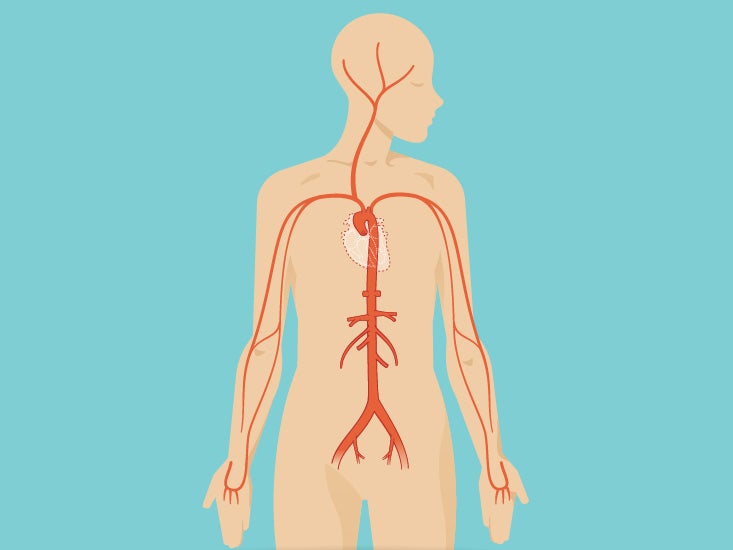
Gsi asked questions about the abdominal membranes to christopher windham, m.d. Unit three — abdominal organs, pelvis & lower limb. Sciency root words make anatomical parts harder to memorize. Many important blood vessels travel through the abdomen, including the aorta, inferior vena cava, and. The anatomy of the liver can be described using two different aspects: Netters posterior abdominal wall labeled chart. But with the use of smart technology, you can learn faster and master abdomen anatomy in no time! Abdominal wall pain clinical evaluation differential. Abdomen anatomy area diagram body maps. A good amount of area is covered by the abdominal wall. Arteries lower leg this mri abdominal arteries anatomy tool is absolutely free to use. Anatomy posters and anatomy charts. This diagram depicts abdominal anatomy.
This diagram depicts picture of abdominal anatomy. Unpaired visceral arteries paired visceral arteries. Sciency root words make anatomical parts harder to memorize. Abdomen anatomy area diagram body maps. • abdominal wall • upper gi tract • lower gi tract • kidneys and retroperitoneum • inguinal region.
Abdomen Anatomy Area Diagram Body Maps from post.greatist.com This abdominal pain diagram and chart defines the meaning of stomach pain using quadrants. Anatomy posters and anatomy charts. Morphological anatomy and functional anatomy. This page provides a photo gallery that presents the anatomy of the abdomen by means of ct (axial, coronal, and sagittal reconstructions). Anatomy of the neuraxis thoracic and abdominal walls. Abdominal wall pain clinical evaluation differential. Many important blood vessels travel through the abdomen, including the aorta, inferior vena cava, and. Diagram showing some of the collateral routes established when portal hypertension exists.
This diagram depicts abdominal anatomy.
Hopefully this provided you with a good overview of the abdominal quadrants, anatomy within each. Abdomen anatomy area diagram body maps. Human anatomy diagrams show internal organs, cells. Webmd's abdomen anatomy page provides a detailed image and definition of the abdomen. Diagram showing some of the collateral routes established when portal hypertension exists. • the abdomen consists of: The abdomen (colloquially called the belly, tummy, midriff or stomach) is the part of the body between the thorax (chest) and pelvis, in humans and in other vertebrates. Learn vocabulary, terms and more with flashcards, games and other study tools. Cystic abdominal masses in children. Netters posterior abdominal wall labeled chart. • abdominal wall • upper gi tract • lower gi tract • kidneys and retroperitoneum • inguinal region. Windham was previously a surgical. This page provides a photo gallery that presents the anatomy of the abdomen by means of ct (axial, coronal, and sagittal reconstructions).
This abdominal pain diagram and chart defines the meaning of stomach pain using quadrants abdominal anatomy. Unit three — abdominal organs, pelvis & lower limb.

